Mole Removal in JVC – Dermatologist-Led Care for Unwanted Moles
A mole, or melanocytic nevus, is a common skin growth formed by pigment-producing cells. Most moles are harmless, but some people seek mole removal in JVC because a mole is noticeable, irritated by clothing or shaving, or has changed in appearance. However, not every mole needs removal, and the right technique depends on its type, size, depth, location, and appearance. At our JVC dermatology clinic, every mole is assessed by a qualified dermatologist before treatment. This careful evaluation helps determine whether removal is appropriate and which approach is most suitable for safe, personalised care.
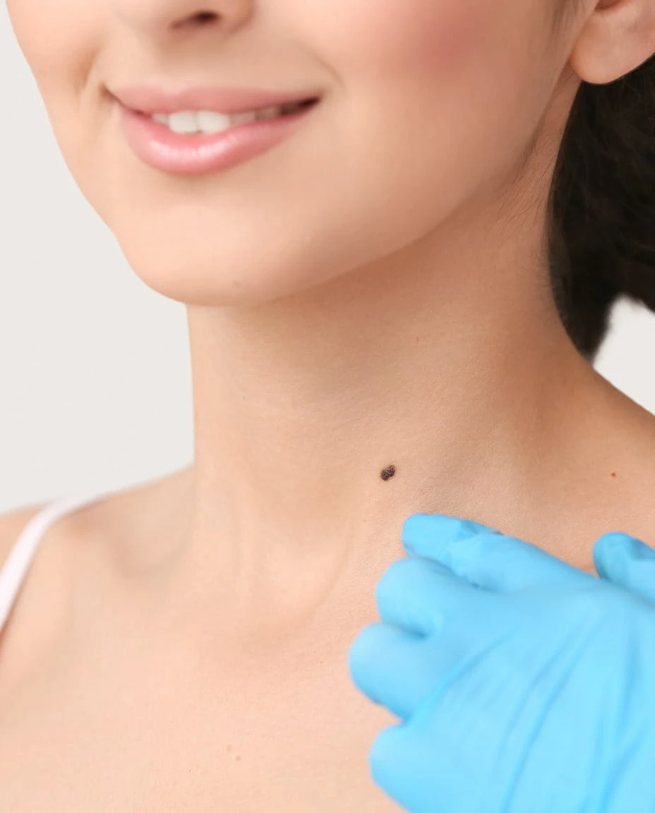
Start With a Mole Assessment
Every mole is different. A professional dermatological assessment helps evaluate its size, shape, colour, location, and recent changes before deciding whether removal is appropriate and which treatment approach is most suitable.
QUICK ANSWER
Mole removal at Vedara Care in JVC, in one paragraph.
Mole removal at Vedara Care begins with a clinical assessment, not a walk-in procedure. A dermatologist examines the mole's shape, colour, border, size, and history before deciding whether it is suitable for removal and, if so, which method is appropriate. Moles that appear stable and benign may be considered for cosmetic removal using techniques such as shave excision or surgical excision, depending on their characteristics. Where a mole shows any feature that raises concern, tissue may be sent for laboratory examination (histopathology) rather than treated as a purely cosmetic matter. Removal technique is never one-size-fits-all — it depends on the individual lesion. Patients should never attempt to remove a mole at home, as this can cause infection, scarring, or delay the diagnosis of a lesion that needed proper evaluation.
WHAT IS A MOLE?
What Is a Mole?
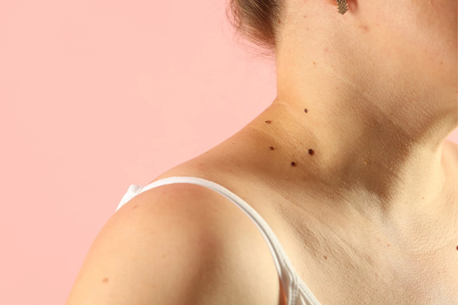
A mole is a common, usually harmless skin growth formed when melanocytes — the cells responsible for skin pigment — cluster together instead of spreading evenly through the skin. Moles can be present from birth (congenital moles) or can develop later in life (acquired moles), often appearing during childhood, adolescence, or pregnancy.
Moles vary considerably in appearance. Some are flat and barely raised above the skin's surface, while others are raised, dome-shaped, or slightly textured. Colour can range from pale brown to dark brown or black, and a person may have moles of several different shades. Most moles remain stable for years and cause no problems at all.
That said, "most moles are benign" does not mean every mole can be ignored indefinitely. Any mole that looks different from a person's other moles, or that changes in a noticeable way, deserves professional attention rather than a guess.
ASSESSMENT
When Should a Mole Be Assessed by a Dermatologist?
Dermatologists commonly use the ABCDE approach as a practical guide for spotting features that warrant closer examination:
A — Asymmetry: One half of the mole looks different from the other half
B — Border: Edges that are irregular, blurred, or notched rather than smooth
C — Colour: Uneven colour, or multiple shades of brown, black, red, or blue within one mole
D — Diameter: Moles larger than roughly 6mm, though smaller lesions can also occasionally be significant
E — Evolving: Any change in size, shape, colour, elevation, or symptoms over time
Beyond ABCDE, other signs that should prompt an assessment include a new mole appearing in adulthood, a mole that bleeds without obvious injury, persistent crusting or ulceration, ongoing itching or pain, and any lesion that is growing noticeably faster than the skin around it.
It's worth being clear: ABCDE is a helpful screening guide, not a diagnostic test. A mole can look unremarkable and still need a closer look, and a mole can tick one of these boxes and still turn out to be entirely benign. The only way to know is a proper clinical assessment — never a home comparison against a photo online.
Why Do Patients Consider Mole Removal?
Patients come to us for mole removal for both cosmetic and clinical reasons, and the two are treated quite differently.
Cosmetic and comfort-related reasons include moles in visible areas such as the face or neck that a patient would simply prefer not to have, moles that repeatedly catch on jewellery or clothing, and moles that make shaving difficult or lead to recurring irritation, nicks, or ingrown hairs. Some patients have lived with a particular mole for years and have simply decided they'd rather not have it anymore — that's a valid and common reason to book a consultation.
Clinically motivated reasons are different in nature. These involve a mole that has changed, looks unusual compared to a patient's other moles, or shows one of the warning signs described above. In these cases, the goal of the appointment isn't cosmetic removal — it's proper assessment, and possibly biopsy.
The distinction matters because it shapes the entire treatment plan. A dermatologist will always clarify, early in the consultation, whether a mole appears to be a straightforward cosmetic case or one that needs further investigation before any removal is considered.
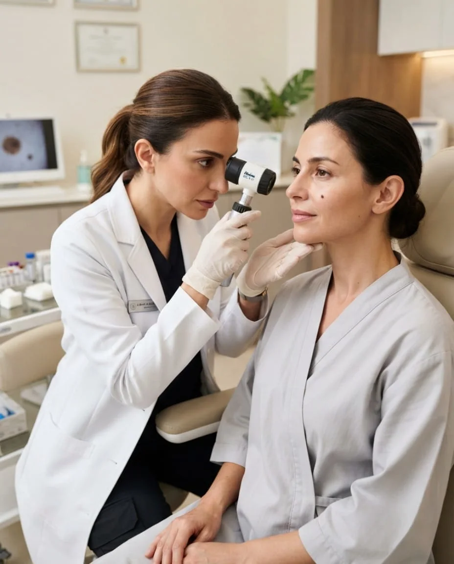
ASSESSMENT BEFORE REMOVAL
Why Mole Assessment Comes Before Removal
No mole should be removed — cosmetically or otherwise — without first being properly assessed. During assessment, the dermatologist will typically consider:
- Visual examination: of the mole's size, shape, colour, and texture
- Medical history: including any personal or family history of skin lesions
- Duration: How long the mole has been present and whether it has changed
- Location: Its exact location and how that may affect treatment choice
- Surface characteristics: such as scaling, crusting, or bleeding
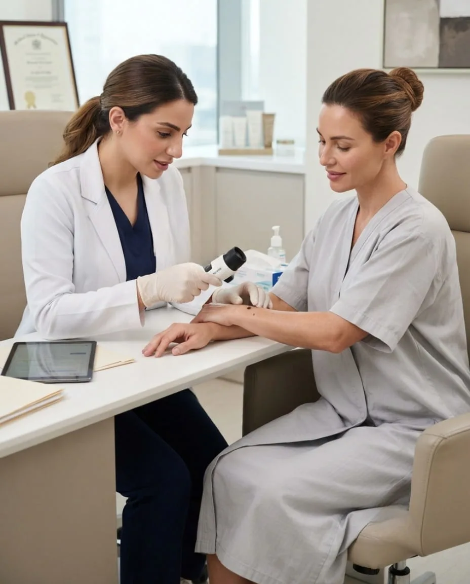
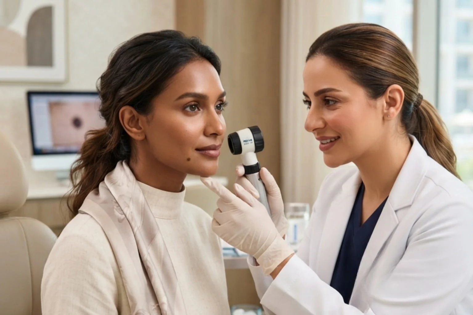
- Dermoscopic assessment: where clinically available, using magnification to examine pigment patterns beneath the skin's surface
- Previous procedures: performed on the same or nearby lesions
This assessment is what allows the dermatologist to decide whether a lesion is appropriate for straightforward cosmetic removal, or whether it needs to be treated with more caution — potentially involving a biopsy before any decision about full removal is made.
TYPES OF MOLES
Types of Moles and Lesions We May Assess
01 — Raised Moles
Raised, dome-shaped moles are often considered for shave excision, though the final decision depends on depth and clinical appearance on examination.
02 — Flat Pigmented Moles
Flat moles can sometimes be more difficult to assess visually and may benefit from dermoscopic examination to understand the pigment pattern before a removal plan is discussed.
03 — Congenital Moles
Moles present since birth are assessed individually, as some congenital moles are larger, deeper, or structurally different from those that develop later in life.
04 — Facial Moles
Moles on the face require particular attention to cosmetic outcome, given the visibility of the area and the importance of scar placement.
05 — Neck and Body Moles
Moles on the neck, torso, or limbs are assessed with consideration for the specific skin tension and healing characteristics of that body area.
06 — Irritated or Frequently Traumatized Moles
Moles that are regularly nicked while shaving, caught on clothing, or otherwise irritated are assessed for both the irritation itself and any secondary changes it may have caused.
07 — Changing or Suspicious Lesions
Any mole showing ABCDE features or other warning signs is assessed with a view toward diagnosis first, and cosmetic considerations second.
Not every lesion identified above is automatically suitable for cosmetic removal — that determination is made individually, after assessment.
REMOVAL METHODS
Which Mole Removal Method May Be Appropriate?
The removal method used depends entirely on what the dermatologist finds during assessment. Common approaches include:
Shave Excision: This involves shaving the raised portion of the mole level with the surrounding skin using a fine surgical blade. It may be considered for certain raised, benign-appearing moles. It can offer a relatively quick recovery, though depending on the depth and nature of the mole, there is a possibility of partial regrowth in some cases.
Surgical Excision: This involves removing the mole along with a margin of surrounding tissue where clinically indicated, followed by sutures to close the wound. It tends to result in a linear scar rather than a rounded mark, and may be the more appropriate option when a fuller tissue sample is needed or when the mole extends deeper into the skin.
Biopsy / Histopathological Examination: In some cases, removed tissue — or a sample of it — is sent to a laboratory for histopathological examination. This is standard practice for any lesion where the clinical appearance raises a question that visual assessment alone cannot answer. A suspicious lesion should never be treated as a purely cosmetic case.
Laser Treatment: Laser treatment is sometimes discussed by patients as an option for mole removal, but it is not appropriate for every pigmented mole. Because laser does not typically provide a tissue sample for laboratory analysis in the same way excision does, it is generally reserved for cases where a dermatologist has already determined, through assessment, that the lesion is suitable for this approach.
IMPORTANT NOTE
Why a Dermatologist Should Assess a Mole Before Laser Removal
This is one of the most important points on this page, so it's worth stating plainly: pigmented lesions often require a diagnosis, not just a removal.
Removing a mole without appropriate prior assessment can interfere with the ability to reach an accurate diagnosis later, particularly if the lesion turns out to have features that needed closer investigation. Laser treatment, in particular, does not provide a tissue specimen for laboratory examination the way excision does — which means that if a mole later needed histopathological review, that opportunity may have already been lost.
For this reason, cosmetic treatment should never be used as a substitute for clinical evaluation. If a mole has any feature that could be considered atypical, biopsy or excision — rather than laser — is generally the more appropriate route, and this decision is one that belongs to the dermatologist, made after assessment, not before.
What Happens During Mole Removal at a Dermatology Clinic?
1. Consultation and Assessment
The process begins with a detailed consultation, including a visual and dermoscopic (where available) examination of the mole, a discussion of the patient's history, and a decision on whether the lesion appears suitable for cosmetic removal or requires further investigation.
2. Treatment Planning
Once assessment is complete, the dermatologist explains the recommended approach, including the technique proposed, what to expect during the procedure, and whether a biopsy or histopathology will be part of the plan.
3. Mole Removal
The chosen technique is carried out under appropriate anaesthesia where required. Procedure time varies depending on the size, depth, and number of moles being treated.
4. Aftercare and Follow-Up
Patients are given wound-care instructions and, where relevant, a follow-up appointment to review healing and discuss any laboratory results.
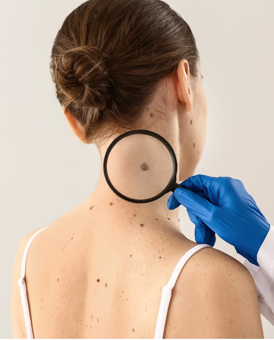
PREPARATION
How Should You Prepare for Mole Removal?
Before your procedure, your dermatologist will want to know about:
- Your general medical history
- Current medications particularly blood-thinning medications
- Any known allergies including reactions to local anaesthetic
- Previous skin infections in the treatment area
- A personal history of keloid or problem scarring
- Recent cosmetic procedures in the same area
- Recent sun exposure or tanning
Please do not stop any prescribed medication before your appointment without first speaking to your prescribing doctor.
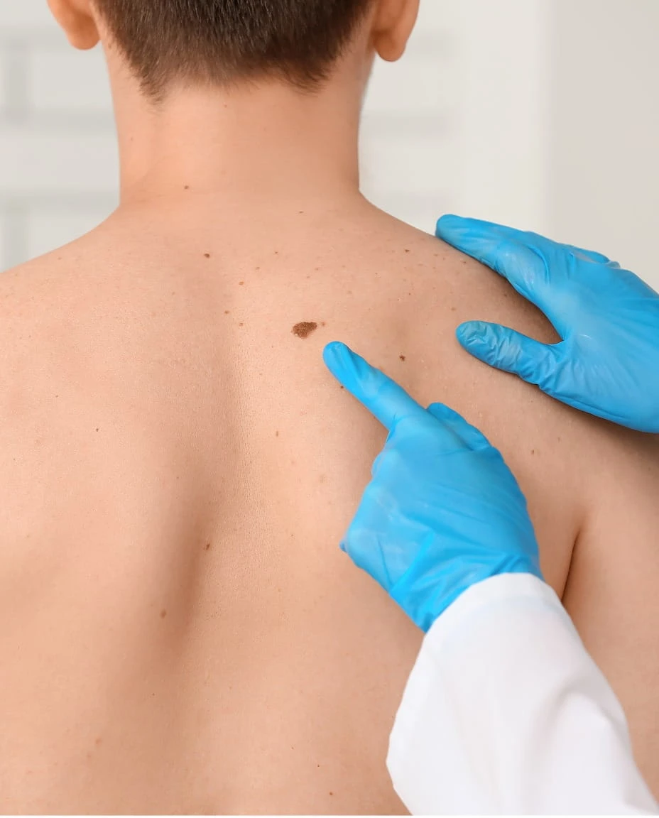
SENSATION
What Does Mole Removal Feel Like?
Sensation during mole removal depends on the technique used. Where local anaesthetic is administered, patients typically feel a brief pinch or sting from the injection, followed by pressure or minor pulling sensations rather than sharp pain during the procedure itself. Procedure duration varies according to the lesion being treated and the method chosen — some appointments are brief, while more involved excisions take longer. No procedure can be guaranteed to be entirely without discomfort, but your dermatologist will discuss what to expect for your specific case.
RECOVERY
What Is Recovery Like After Mole Removal?
Immediately after removal: The area may be covered with a dressing. Mild bleeding or oozing in the first few hours is not unusual.
First 24–48 hours: Some redness, tenderness, and mild swelling around the site is common.
First week: A scab or crust typically forms as part of normal healing. It's important not to pick at this, as doing so can affect the final scar.
Following weeks: The area continues to heal and settle. Temporary sensitivity or changes in colour around the site can occur during this period.
Recovery timelines vary considerably depending on the removal method, the mole's location, and individual healing.
SCARRING
Will Mole Removal Leave a Scar?
Any procedure that removes tissue from the skin carries the possibility of leaving a mark or scar — this is simply a fact of how skin heals, and no reputable clinic should promise otherwise. How noticeable a scar becomes depends on several factors: the removal method used, the size and depth of the original mole, the location on the body, and each individual's healing tendencies. Skin type can also influence whether temporary pigment changes occur around a healed area.
Proper aftercare supports the healing process, but it cannot override biology. The honest goal of treatment is this: "the goal is to achieve the best possible cosmetic outcome while treating the lesion appropriately" — not to promise an outcome that cannot be guaranteed.
EXPECTATIONS
What Should You Expect After Mole Removal?
Once the chosen technique has been carried out, the unwanted mole is removed, and the treated area begins the normal healing process. In the early stages, some redness or a visible wound is expected — this is not a sign that anything has gone wrong. Healing occurs gradually over the following weeks, and scar maturation can continue for months after the procedure. Temporary pigment changes around the area are possible during this time. As with any procedure, the final appearance should only be judged once healing is reasonably complete, not in the first days or weeks.
BEFORE AND AFTER
Mole Removal Before and After: What Should Patients Understand?
Before-and-after photographs can be a useful way to understand what mole removal can achieve, but they should be read with some context. Results vary depending on the individual mole, and photographs can be affected by lighting, camera angle, and the stage of healing shown. An early photograph taken shortly after treatment may show redness or a visible wound that will continue to fade and settle over time — this is normal and does not reflect the final result. Any before-and-after images shared by Vedara Care are real, consented patient photographs; we do not use stock or illustrative images to represent clinical outcomes.
CANDIDATES
Who May Be a Suitable Candidate for Mole Removal?
Mole removal may be appropriate for patients who:
- Have had their mole assessed and found to appear benign
- Want removal for cosmetic reasons
- Experience repeated irritation from a mole in a specific location
- Have a mole affected by shaving or friction from clothing
- Have a lesion in a position that is inconvenient or bothersome
- Have received a specific removal recommendation from a dermatologist following assessment
Suitability is always determined individually, after a full examination — not from a description or photograph alone.
INVESTIGATION
When Should a Mole Be Investigated Rather Than Simply Removed Cosmetically?
Some moles need to be treated as a diagnostic question before they can be treated as a cosmetic one. This includes moles that are changing in size, shape, or colour; lesions with asymmetrical shape or irregular, blurred borders; moles with multiple colours or shades; lesions growing noticeably faster than expected; and any mole that bleeds, ulcerates, or develops persistent crusting without an obvious cause.
In these situations, biopsy or full excision with histopathological examination may be the more appropriate route, rather than a technique chosen primarily for cosmetic reasons. We are not able to diagnose any skin condition, including melanoma or other skin cancers, from an online description or photograph — this can only be determined through in-person clinical assessment and, where indicated, laboratory examination.
SAFETY & SIDE EFFECTS
Mole Removal Safety and Possible Side Effects
As with any skin procedure, mole removal carries some possible risks, which vary depending on the technique used:
- Bleeding: usually minor and short-lived
- Infection: at the treatment site
- Pain or tenderness: during healing
- Swelling: around the treated area
- Scarring: ranging from minimal to more noticeable
- Pigment changes: Temporary or, less commonly, longer-lasting
- Delayed healing: particularly in certain skin types or health conditions
- Recurrence: or partial regrowth of the mole in some cases, depending on technique
- Allergic reaction: Rarely, to local anaesthetic
- Additional treatment: The possibility that additional treatment may be recommended depending on laboratory findings
SKIN TYPES
Mole Removal and Darker Skin Tones
Patients with darker skin tones can experience specific healing considerations after mole removal, including a higher likelihood of post-inflammatory hyperpigmentation (darkening of the healed area) or, less commonly, hypopigmentation (lightening). Scar pigmentation can also differ from lighter skin types. Sun protection during healing plays an important role in minimising these changes, and treatment technique is selected with these factors in mind. No single removal method is universally the safest choice for all skin types — this is part of what makes individual assessment so important.
FACIAL MOLES
Mole Removal on the Face: Why Technique Matters
Facial moles require particular care because of how visible the area is and how much cosmetic outcome matters to patients. Considerations include scar placement in relation to natural facial lines, skin tension across different parts of the face, and the healing characteristics of facial skin compared with the body. Dermatologists generally favour more conservative techniques on the face where clinically appropriate, precisely because the cosmetic stakes are higher. This is why facial mole removal, more than almost any other area, benefits from a dermatologist's individual assessment rather than a standard, one-size-fits-all approach.
AFTERCARE
How to Care for the Area After Mole Removal
General aftercare principles include:
- Keep the treated area clean following your dermatologist's specific instructions
- Follow all wound-care guidance provided at your appointment
- Do not pick at scabs or crusting
- Avoid unnecessary friction or pressure on the area
- Follow any dressing-change instructions exactly as given
- Use prescribed medication or ointments only as directed
- Protect healed skin from sun exposure particularly in the months following treatment
- Attend any recommended follow-up appointment
LABORATORY EXAMINATION
Why Some Removed Moles Are Sent For Laboratory Examination
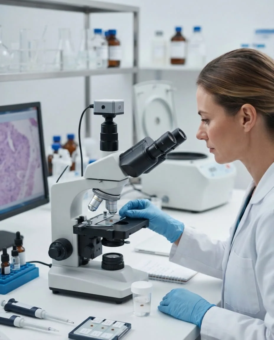
Histopathology is the microscopic examination of tissue by a laboratory specialist to determine its exact cellular composition. When a mole is removed, tissue may be sent for this examination — not because something is necessarily wrong, but because visual appearance alone cannot always establish a complete diagnosis, even for an experienced dermatologist.
Laboratory analysis provides a level of certainty that examination by eye cannot. It confirms whether a lesion is benign, and if any atypical features are found, it allows for appropriate next steps to be planned. If you are unsure whether your removed tissue will be examined, it is entirely appropriate to ask your dermatologist directly during your consultation.
PRICING
What Does Mole Removal Cost in JVC?
| Service | Price |
|---|---|
| Dermatology consultation | AED[X] |
| Mole assessment | AED[X] |
| Mole removal | AED[X] |
| Additional lesion removal | AED[X] |
| Histopathology, where required | AED[X] |
| Follow-up consultation | AED[X] |
The overall cost of mole removal in JVC can depend on several factors, including the number of moles being treated, their size, location on the body, the removal technique selected, the complexity of the case, whether anaesthesia is required, whether histopathology is needed, and any follow-up appointments included in your treatment plan.
Frequently Asked Questions
Short, honest answers from our clinical team. Cannot find what you are looking for? WhatsApp us, usually answered in under 15 minutes.
Ask a QuestionVISIT OUR JVC CLINIC
Mole Assessment and Removal at Our JVC Dermatology Clinic
NEARBY LANDMARKS
- ✓Walking distance from Circle Mall
- ✓3 min from FIVE Jumeirah Village Hotel
- ✓5 min from JSS Private School
- ✓Free patient parking
DISTANCE FROM DUBAI COMMUNITIES
Important Information
Medical Disclaimer
This page is for informational purposes only and does not constitute medical advice. All treatments carry risks and benefits that should be discussed with a qualified healthcare professional. Results vary between individuals. Vedara Care Polyclinic is a DHA-licensed facility.
READY TO START?
Start With a Dermatologist-Led Mole Assessment
Whether you're considering removal of a mole that's bothered you for years, or you've noticed a change that's made you want it checked, the right first step is the same: a proper assessment. Your dermatologist will look at the mole's appearance, how it has changed over time, its location, size, and your skin type, along with your broader medical history — before recommending an appropriate removal technique, discussing whether histopathology may be needed, and explaining what to expect during healing, including the realistic possibility of scarring.
RELATED SERVICES
Related Dermatology Services and Resources
Dermatology in JVC
Explore personalised dermatology care for a wide range of skin concerns, with treatment plans based on your skin type and individual needs.
Learn moreSkin Diseases
Get professional assessment and personalised care for common and ongoing skin concerns affecting the face and body.
Learn morePigmentation Treatment
Learn about treatment options designed to improve the appearance of uneven skin tone, dark spots, and pigmentation concerns.
Learn moreScar Treatment
Discover personalised approaches for reducing the appearance of acne scars, surgical scars, and other types of skin scarring.
Learn moreSkin Rejuvenation
Laser skin treatment to remove scarring and improve skin textureExplore non-surgical treatments that can help refresh the skin, improve texture, and support a healthier-looking appearance.
Learn more